
With any type of surgery, precision is essential. Accuracy seems even more crucial when the area of concern is the patient’s face. When evaluating a patient for oral surgery, doctors need the most advanced imaging tools available to provide a detailed diagnosis that will enable them to come up with an effective treatment plan. A 3D imaging system produces high-definition panoramic images to allow the oral surgeons to accurately diagnose the issue at hand, effectively explain the situation to the patient, and efficiently address the problem. The use of 3D imaging is substantially beneficial when assessing oral surgery-related issues for several reasons, as explained below.
What is 3D Imaging?
When it comes to oral and facial imaging, a 3D scan allows doctors, dentists, orthodontists, oral surgeons, and others to view the facial bones and teeth in superb detail on a computer screen that can be adjusted to allow views from any angle.
The 3D imaging system produces thousands of scans that combine on the screen to create a movable and transferable digital image that makes it easier to evaluate the patient’s issue, offer solutions, and share images with another consulting doctor.
What Kind of Technology is Used for the Most Effective 3D Imaging?
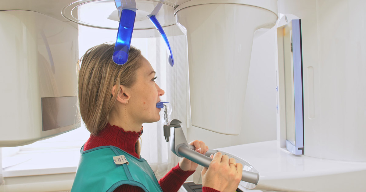
The most advanced 3D imaging systems use cone beam computed tomography (CBCT) technology, which allows doctors to capture a digital image of the patient’s teeth and bone structure to assist with creating a detailed treatment plan using a computerized visual model that can be viewed in 3D from any angle.
The image is captured by a trained technician using a low dose of radiation to create a clear scan of the area to be addressed.
The cone beam allows the imaging to be quickly performed while limiting the scan to the area of concern, reducing the need to expose patients to unnecessary amounts of radiation.
What are the Benefits of 3D Imaging?
Doctors and patients benefit from the following advantages of 3D imaging:
- It improves diagnostics. Clear and moveable 3D scans give doctors the ability to assess the problem with sharp visibility from any angle.
- It informs patients. Useful visuals allow patients to gain a clear understanding of the issues to be addressed.
- It is easily shared. The digital images can be shared effortlessly with the referring doctors.
- It minimizes risks. 3D imaging is quicker and more targeted than traditional scans, minimizing the patient’s exposure to radiation.
- It is convenient. In-house scanning allows doctors to offer imaging and procedures in the same visit.
Mullica Hill Oral Surgeons at Lanzi Burke Oral & Maxillofacial Surgeons Use the Latest Technology to Serve Patients
Patients in need of oral surgery want to know that they are getting the best care using the most advanced technology and techniques. The use of 3D imaging allows doctors to get a clear picture of the problem and implement ways to address the issue. At the same time, the innovative imaging provides a clear way for patients to understand their unique issues and make informed decisions about their care. The Mullica Hill oral surgeons at Lanzi Burke Oral & Maxillofacial Surgeons use the latest 3D imaging technology to assess the specific issues at hand and create a custom treatment plan to address the patient’s needs. Call us at 856-582-4222 or contact us online to make an appointment or schedule a consultation. With offices in Washington Township, Haddonfield, and Woolwich Township, New Jersey, we are dedicated to helping patients throughout South Jersey.